Miter
Lilliputian-sized, you
are a gathering of bishops
under cathedrals of oaks.
I kneel at your feet…
From Light and Matter 2008


Get Tips: Nature Photography
Once a month a new segment will be sent to you on trying new techniques, using certain gear, stalking certain species or working in post-processing. Learn how to take stunning photographs of wildlife and landscapes by checking out the photo tips section.


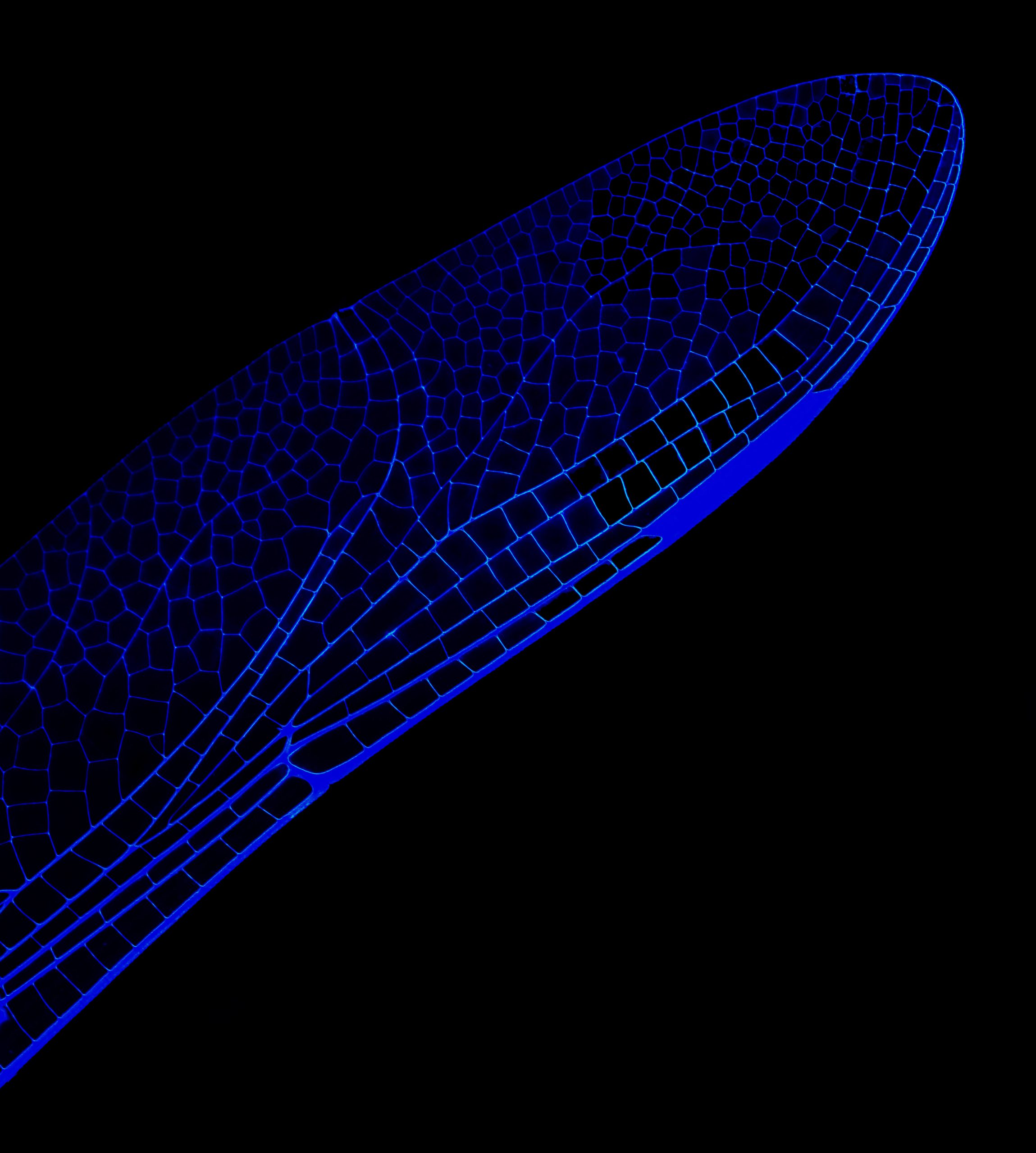
Scanning Electron Microscope Images
Several years ago, I decided I wanted to learn how to do SEM (Scanning Electron Microscope) photography. I wrote a grant and a year later I took my first round of images. The amazing part of this process is the ability to go deeply into any sample, up to 100,000 times magnification! I mostly stay within the 25x -650x magnification range so the specimens are identifiable.
The process is complicated and it took some time for me to understand how to get the images in focus appropriately. This is very different than a normal photograph as there is a directed beam of electrons that is focused on an area of the specimen (say a honeybee for example). There can be issues if there is not enough air taken away or the sample is in the wrong position. And the beam can actually move certain parts of the sample (even though it is not alive) like it is being swayed by wind!
In the end, I want to see the world differently and to show viewers an image they have never seen before. That is what the best of photography does; it pushes the boundaries and opens up new possibilities of knowing.


